

The human heart begins to form very early on during embryonic development. Step by step, a few precursor cells give rise to a highly complex organ with various chambers, valves, blood vessels, and a specialized heart muscle. For this process to work, cells must constantly communicate with one another and respond to signals from their environment. Researchers at the University of Copenhagen have now identified a previously undescribed mechanism involved in this communication. At the center of this process is a microscopic structure that protrudes from the surface of cells: the so-called primary cilium.
The scientists discovered that several proteins assemble there to form a kind of molecular switch. If this system is disrupted by certain genetic mutations, it can impair the development of the heart. The findings thus provide a possible new explanation for some congenital heart defects—particularly those in which other organs are also affected. The study was published in the journal PLOS Biology.
When the Development of the Heart Becomes Unbalanced While Still in the Womb
Congenital heart defects do not develop only after birth. The structural changes form during embryonic development. A wide variety of abnormalities can occur. In some children, for example, the septum between the ventricles or atria is not fully formed. In others, heart valves, major blood vessels, or the shape and connections of individual heart chambers are affected.
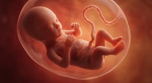
The severity of such a defect can therefore vary greatly. Some abnormalities are not detected until years later, while severe forms require treatment immediately after birth.
Congenital heart defects are among the most common birth defects worldwide. According to the World Heart Federation, approximately 1.4 to 2.3 percent of all children born worldwide are affected. For the year 2023, the number of affected newborns is estimated at about 2.3 million. At the same time, an estimated 16 million people worldwide were living with a congenital heart defect. Precisely because these conditions are so diverse, there is no single cause.
In some affected individuals, known genetic mutations play a role. In other cases, the interplay between genes and the numerous molecular processes that occur during embryonic development remains largely unclear. The new study addresses precisely this issue.
A Tiny “Antenna” Monitors the Cell’s Environment
The researchers focused on the primary cilium. This is a microscopic, antenna-like structure located on the surface of many cells. Unlike motile cilia, which can transport fluids or mucus, for example, the primary cilium primarily serves as a sensor and communication platform. The structure can detect signals from the cell’s surroundings. These include, among other things, signals from growth factors and other signaling molecules.
This information is crucial for a developing cell. It must respond to the signals and adjust its behavior accordingly. Depending on the situation, such a signal can, for example, cause the cell to divide, alter its properties, move, or differentiate into a specific cell type. During embryonic development, this communication becomes particularly important. This is because, during this phase, highly complex organs must emerge from what are initially relatively simple cell clusters.
In this process, the primary cilium acts, in a sense, as an interface between the cell’s external environment and the molecular processes taking place inside. The University of Copenhagen therefore vividly describes it as a kind of “antenna” for the cell.
Three Proteins Form a Key Signaling Hub
The new study now shows that the primary cilium hosts a specific signaling pathway during heart development. The focus is on the proteins TAK1, TAB2, and PKA-Cα. In the scientific paper, TAK1 is also referred to by its gene/protein name MAP3K7, while PKA-Cα is encoded by the PRKACA gene. The three components interact within the primary cilium.

This involves what is known as a non-canonical TGFB/BMP signaling pathway. TGFB and BMP signals are already known to be important regulators of the development of various tissues. The new study shows that TAK1 and its upstream regulators, TAB2 and PKA-Cα, also play a role directly at the primary cilium. This signaling pathway is activated during the development of cardiac muscle cells. This activation can be amplified by TGFB and BMP signals.
This paints a new picture of how a cell responds to signals during development: It is not only the known signaling pathways within the cell that are crucial. The location where a signal is received and processed can also play an important role.
What Happens When TAK1 is Absent?
To determine the actual significance of this mechanism, the researchers modified the relevant genes in various experimental models. One of these was zebrafish. Zebrafish are particularly useful for developmental research because their early development can be easily observed. The scientists generated fish in which tak1 or tab2 was knocked out.
The consequences were clear: Among other things, the modified fish developed abnormalities in the heart. In TAK1-deficient zebrafish, the researchers also found alterations in gene networks associated with the development of the heart muscle, the extracellular matrix, and other components of the heart.
In TAB2 mutants, changes in heart structure and heart function were also observed. Among other things, the experiments revealed abnormalities in the heart’s ventricles, ejection fraction, and trabeculation.
The results thus provided strong evidence that the proteins under investigation are not merely present in the primary cilium by chance. They do indeed appear to be relevant to cardiac developmental processes.
The Researchers also Identified the Mechanism in Human Cells
However, the study was not limited to fish. The research team used various cell models and examined, among other things, human cells as well as stem cell models capable of differentiating into cardiomyocytes.
In the process, the researchers observed that TAK1 is localized to the primary cilium of cells. TAB2 and PKA-Cα were also detected in this structure. In cardiac progenitor cells, the connection between the primary cilium and the signaling pathway under investigation was also evident.
The result: Signal transmission at the primary cilium is impaired, and differentiation into cardiac muscle cells is inhibited. This allowed the researchers to understand the connection between the tiny cellular structure and the development of the heart muscle at the cellular level as well.
Genes from People With Heart Defects also Provide Clues
Another part of the study led the researchers directly to patients with congenital heart defects. The team analyzed genetic data from several thousand people with congenital heart defects and searched for rare variants in genes responsible for components of the signaling pathway under investigation.

Particularly striking was an increased prevalence of rare variants in TAB2 and TAK1 among people with congenital heart defects who also had abnormalities outside the heart. The researchers therefore focused specifically on so-called syndromic congenital heart defects.
The original study cites an analysis of 3,876 people with congenital heart defects compared to 45,082 control subjects. However, this genetic data alone would not have been sufficient to prove that the variations are actually responsible for the heart defects. That is why the next step was crucial: The scientists investigated the biological consequences of the relevant variants.
Why Some Genetic Disorders Affect Multiple Organs
For certain patient-specific TAK1 variants, the researchers observed an altered localization of the protein. This is biologically significant, because a signaling protein can only function properly if it is in the right place. If TAK1 no longer reliably reaches the primary cilium, the signal transduction occurring there can be disrupted.
The study’s findings therefore suggest that it is not only the quantity of a protein that matters. Its spatial position within the cell can also play an important role in heart development. This is one of the most interesting aspects of the study.
The discovery could also explain why certain genetic disorders affect more than just the heart. The genetic mutations studied are associated with so-called syndromic disorders. In these disorders, a congenital heart defect occurs alongside abnormalities in other organs or tissues. This aligns with an important characteristic of the primary cilium: it is present in a wide variety of cell types.
If a fundamental mechanism of this cellular structure is disrupted, it could therefore have effects in multiple parts of the body. In the zebrafish experiments as well, the researchers did not find changes exclusively in the heart. In the genetically modified fish, abnormalities were observed in structures outside the heart, among other things. TAK1 was also detected in cilia from tissues outside the heart. The scientists see a possible connection here with the fact that some patients simultaneously develop changes in the heart, brain, kidneys, or skeleton.
This discovery could therefore have implications beyond the study of congenital heart defects. Disorders of ciliary function are already associated with numerous rare genetic diseases. However, exactly what happens at the molecular level is not known in every case. The mechanism now described could provide a missing link.
If scientists gain a better understanding of which proteins interact in the primary cilium and how these signaling pathways are regulated during embryonic development, it might also be possible to better explain other diseases. Søren Tvorup Christensen of the University of Copenhagen sees this as a potential common mechanism for diseases that have so far been difficult to link to one another.
No New Treatment Yet for Affected Children
As interesting as the results are, they do not yet lead to a new therapy for congenital heart defects. The researchers have identified a biological mechanism and investigated it using several experimental approaches. However, a large portion of the key findings comes from cell and animal models.
This means that while the scientists have strong evidence suggesting that the mechanism is also relevant in humans, they cannot yet directly demonstrate every detail of the processes occurring in the human embryo.
The University of Copenhagen also explicitly points out that, due to its genetic analyses and experimental models, the study cannot definitively prove the mechanism in humans. However, the results as a whole provide strong evidence of its significance.
An Important Step from Gene to Disease
The true significance of the work may therefore lie in the fact that it establishes a connection between different levels. It begins with rare genetic mutations in people with congenital disorders. Experiments then show that corresponding mutations in model organisms can cause developmental abnormalities. Finally, cellular studies explain which molecular signaling pathways might be disrupted in the process.

This creates a biological chain: Genetic mutation → disrupted signaling pathway in the primary cilium → altered development of heart muscle cells → possible heart malformation. Of course, reality is more complex than this simplified representation. Congenital heart defects have numerous different causes, and not every heart defect is likely attributable to this signaling pathway. However, the new study provides another building block that can help us understand the development of certain forms of the disease.
A Tiny Structure with Far-Reaching Significance
The idea that a structure so small it can only be seen under a microscope—located on the surface of a cell—can influence the development of an entire organ makes the significance of this discovery particularly striking.
The primary cilium is only a few micrometers in size. Nevertheless, it can receive information from a cell’s surroundings and thereby influence processes that determine the characteristics the cell will later possess.
This communication is particularly critical during embryonic development. Even small changes in a signaling pathway can trigger a cascade of further changes. In heart development, this is precisely what could happen if TAK1, TAB2, or PKA-Cα do not function properly or fail to reach their intended location in the primary cilium.
A New Piece of the Puzzle in the Development of Congenital Heart Defects
The next steps will therefore involve investigating the mechanism in even greater detail. Among other things, this involves determining which genetic changes have particularly severe consequences and why some variants primarily affect the heart, while others also affect the brain, kidneys, or skeleton.
Equally important is the question of whether the findings can eventually be applied to medical diagnostics. Lars Allan Larsen and his colleagues hope that a better understanding of ciliary signaling pathways could, in the long term, help detect certain rare genetic disorders earlier. The development of targeted treatments could also benefit from this one day. Until then, the work remains an important contribution to basic research.
Congenital heart defects do not result from a single error in a single biological process. They can stem from a wide variety of genetic and developmental causes. However, the study from the University of Copenhagen now shows that a previously poorly understood communication platform within the primary cilium plays an important role in heart development.
TAK1, TAB2, and PKA-Cα form a signaling pathway there that is active during the development of heart muscle cells. Genetic alterations can disrupt this process. Experiments on zebrafish and cell models show that such disruptions can be associated with changes in heart development and, in some cases, the development of other tissues as well.
The researchers have thus revealed another link between genes, cell communication, and the onset of congenital disorders. This does not yet explain all congenital heart defects. But the tiny “antenna” on the surface of our cells could turn out to be a surprisingly important part of the biological system that determines how the heart develops even before birth.
Related Posts
-

Congenital heart defects can have a major impact upon pregnancy. Therefore, it is important that…
-

Heart palpitations that occur during pregnancy are typically not a cause for concern and can…
-

Although it may increase risk factors, being stricken with heart disease does not necessarily mean…


