
Preeclampsia is one of the most complex conditions of pregnancy and is a leading cause of maternal and fetal morbidity worldwide. Clinically, it is traditionally defined by the onset of hypertension after the 20th week of pregnancy, often accompanied by proteinuria, edema, and, in severe cases, organ dysfunction such as liver or kidney involvement.
For a long time, preeclampsia was understood primarily as a hypertensive disorder of pregnancy—that is, as a problem with blood pressure regulation in the third trimester. This classical model was heavily symptom-oriented and based on the clinical observation of hypertension, proteinuria, and edema as the key symptoms. For many years, the underlying mechanisms remained only partially understood and were often interpreted primarily in the context of maternal dysregulation of the cardiovascular system. This understanding has changed fundamentally in recent years.
Preeclampsia: More Than Just High Blood Pressure
Advances in placental research, molecular vascular biology, and biomarker diagnostics have shown that preeclampsia develops much earlier in the course of pregnancy and is much more strongly influenced by placental developmental processes than previously assumed. Modern research now increasingly describes preeclampsia as a systemic, placenta-triggered vascular disease in which the mother’s clinical symptoms are understood not as the primary pathological process, but as a secondary consequence of early abnormal development of placental perfusion and signal regulation.

The focus is not primarily on the cardiovascular system itself as the point of origin of the disease, but rather on the placenta as the initial pathophysiological organ. It functions not only as an organ for supply and exchange between mother and fetus, but also as a highly active endocrine and immunological signaling center. In cases of impaired development—particularly with inadequate remodeling of the uterine spiral arteries and the resulting hypoxic stress—the secretory profile of the placenta undergoes profound changes. This leads to the increased release of factors that act systemically on the maternal vascular system, triggering widespread endothelial dysfunction there.
This biochemical communication allows preeclampsia to be understood as a biphasic process: an early, often clinically silent phase of placental maldevelopment and a later, systemic phase in which the maternal vascular response dominates. In this model, local changes in the placenta—such as hypoxia, impaired trophoblast invasion, and altered angiogenesis—interact directly with generalized vascular effects throughout the mother’s entire body. The resulting clinical picture is thus neither purely placental nor purely maternal cardiovascular, but arises from the dynamic interaction of both systems.
In this way, the understanding of preeclampsia shifts away from an isolated hypertensive disorder toward an integrated model of placenta-induced systemic endothelialopathy, in which the mother’s vascular function serves as the target structure of dysregulated placental signaling.
Early Pregnancy as a Critical Phase in Disease Onset
Absent Remodeling of the Spiral Arteries
In many cases, the origins of preeclampsia lie very early in pregnancy, often as early as the first trimester, long before clinical symptoms appear. Normally, a profound remodeling of the uterine spiral arteries occurs during this phase. These vessels are altered by trophoblast cells in such a way that they lose their original muscular and highly regulatory character and instead become wide-lumen, low-resistance vessels.
This remodeling is essential to ensure adequate and stable blood flow to the placenta. In preeclampsia, however, this process is often incomplete or disrupted. The spiral arteries partially retain their high resistance, leading to reduced and pulsatile placental perfusion. This hemodynamic instability is one of the central triggers of the subsequent disease cascade.
Placental Hypoxia as a State of Biological Stress
Due to the restricted blood flow, a state of relative oxygen deprivation develops in the placenta. This hypoxia is not a short-term event but a chronic stimulus that triggers profound changes in placental biology.
Cells respond to this oxygen deficiency by activating specific transcriptional programs, particularly hypoxia-inducible factors such as HIF-1α. These signals alter the expression of numerous genes involved in angiogenesis, cell growth, and metabolism.
The placenta initially attempts to compensate for this insufficient supply. At the same time, however, factors are also produced that act far beyond the placenta and influence the maternal vascular system.
Antiangiogenic Imbalance: Disruption of Vascular Communication
sFlt-1 and PlGF as Key Mediators
A crucial mechanism in the pathophysiology of preeclampsia is the imbalance between proangiogenic and antiangiogenic factors. The ratio of soluble fms-like tyrosine kinase-1 (sFlt-1) to placental growth factor (PlGF) has been particularly well studied.
In preeclampsia, there is increased release of sFlt-1 from the hypoxic placenta. This molecule binds circulating proangiogenic factors such as PlGF and VEGF, thereby reducing their biological availability. At the same time, the concentration of functionally active growth factors decreases.
The result is a systemic antiangiogenic environment that significantly impairs the normal function and regeneration of the vascular system.
Consequences for the Mother’s Vascular System
This biochemical shift directly affects the mother’s vascular physiology. Endothelial cells, which normally play a central role in regulating vascular tone and blood flow, increasingly lose their functional stability.
Typical changes include reduced production of nitric oxide (NO), one of the body’s most important vasodilatory signaling molecules. At the same time, sensitivity to vasoconstrictive stimuli increases.
In addition, the balance between procoagulant and anticoagulant factors shifts, which can lead to a prothrombotic environment. This combination of vasoconstriction, increased vascular stiffness, and impaired microcirculation explains many of the clinical symptoms of the disease.
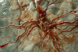
Endothelial Dysfunction as a Systemic Disease Process
The Endothelium as a Central Target Organ
The endothelium is far more than just a passive lining of the blood vessels. It functions as a highly active endocrine organ that continuously produces signals to regulate vascular diameter, inflammatory status, and hemostatic balance.
In preeclampsia, the endothelium is systemically activated and becomes dysfunctional due to circulating placental factors. This endothelial dysfunction is not locally confined but affects nearly all vascular regions of the body.
The consequences are wide-ranging: increased peripheral resistance, impaired organ perfusion, and increased sensitivity to inflammatory stimuli.
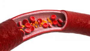
The Role of the Endothelial Glycocalyx
An increasingly important area of research is the endothelial glycocalyx, a fine, gel-like structure composed of proteoglycans and glycoproteins that covers the luminal surface of blood vessels. This structure acts as a protective barrier and plays a central role in mechanical and biochemical signal transduction between the blood and the vascular wall.
In preeclampsia, studies suggest damage to or degradation of this glycocalyx layer. This leads to increased vascular wall permeability, enhanced leukocyte adhesion, and impaired regulation of microcirculation.
The loss of the glycocalyx is increasingly viewed as a key mechanism that exacerbates endothelial dysfunction and could represent a therapeutic target in the future.
Inflammation and Oxidative Stress as Pathology-Exacerbating Factors
Secondary but Crucial Mechanisms

In addition to antiangiogenic imbalance, inflammatory processes and oxidative stress play an important role in the progression of preeclampsia. These mechanisms are often triggered by initial placental hypoxia and exacerbate preexisting vascular dysfunction.
Oxidative stress arises from an imbalance between the production of reactive oxygen species and the body’s antioxidant defense mechanisms. In the placenta and the maternal circulatory system, this leads to further damage to endothelial cells and an amplification of inflammatory signaling cascades.
Context within the Disease Model
Current research, however, emphasizes that oxidative stress should not be understood as the primary cause. Rather, it acts as a secondary amplifying mechanism that stabilizes and exacerbates the preexisting pathophysiological cascade.
This perspective has important implications for therapeutic approaches, as purely antioxidant strategies have so far shown only limited success in clinical trials.
Extracellular Vesicles as a Communication System Between the Placenta and the Mother
Biological Signal Carriers with Systemic Effects
Another area of intensive research is extracellular vesicles, small membrane-enclosed particles released by the placenta into the maternal bloodstream. These vesicles contain a variety of biologically active molecules, including proteins, lipids, and RNA fragments.
They function as highly specialized communication systems between the placenta and the maternal organism and can transmit targeted signals to distant target cells.
Changes in Preeclampsia
In preeclampsia, there is both an increased release and an altered composition of these vesicles. The signal molecules they contain promote inflammatory processes, activate endothelial cells, and reinforce the antiangiogenic environment.
This creates a self-reinforcing cycle in which placental signals further drive systemic vascular changes, which in turn exacerbate placental dysfunction.
Clinical Significance and Long-Term Consequences
Preeclampsia as a Marker of Vascular Vulnerability
Preeclampsia is increasingly viewed not only as an acute pregnancy-related condition but also as an indicator of a long-term cardiovascular risk profile. Epidemiological studies show that women who develop preeclampsia during pregnancy have an increased risk of developing hypertension, cardiovascular system, and stroke later in life.
These observations lead to the hypothesis that preeclampsia acts as a kind of “stress test” for the maternal vascular system, during which latent weaknesses become clinically apparent for the first time.
New Therapeutic Approaches in Vascular Research
Shifting from Symptom Control to Treating the Underlying Causes
The standard treatment for preeclampsia is based on blood pressure control and close monitoring of both mother and child. Since causal treatment is currently only possible through delivery, a major focus of current research is on developing new approaches that intervene earlier in the pathophysiology.
The focus is on strategies that act at the molecular level: the modulation of antiangiogenic factors, the stabilization of endothelial function, and the protection of structural components such as the glycocalyx.
In the long term, research aims not only to detect and monitor preeclampsia but also to actively intervene in its development. This includes biomarker-based early diagnosis, individualized risk profiles, and potential therapeutic interventions in early pregnancy. These developments could fundamentally transform the treatment of preeclampsia and help significantly reduce both maternal and fetal risks.
Conclusion: A Paradigm Shift in Understanding the Disease
Preeclampsia research is undergoing a profound transformation—moving away from a purely symptom-oriented understanding toward a complex model of a placenta-triggered systemic vascular disease.
The integration of placental biology, endothelial research, and molecular signal transduction opens up new diagnostic and therapeutic possibilities and helps to detect the disease earlier and treat it more effectively in the future.
Related Posts
-

Ptyalism is a condition that causes the overproduction of spit. Salivation, generally, is incredibly useful.…
-

An analysis of cheek swabs from pregnant women has revealed a potential epigenetic biomarker for…
-

What is Preeclampsia? Previously referred to as toxemia, preeclampsia is a medical condition that affects…