

Pregnancy can leave traces that extend far beyond childbirth. Scientists have discovered that individual cells from the fetus enter the mother’s body and can still be detected there decades later. This phenomenon is known as fetal microchimerism. Researchers are now investigating whether these cells interact with the immune system, are involved in repair processes, or may even play a role in certain diseases. But much remains unclear.
When the Child’s Cells Remain in the Mother’s Body
During pregnancy, an intense biological connection develops between mother and child. Oxygen and nutrients are transported via the placenta, metabolic waste products are excreted, and numerous signals are exchanged between the two organisms. Another process takes place here that has long received less attention: Individual cells can cross the placenta and enter the other’s bloodstream.
This exchange occurs in both directions. Not only can fetal cells enter the mother’s body, but maternal cells can also enter the fetus’s body. Some of the cells involved can subsequently be detected in various tissues. It is particularly noteworthy that some fetal cells can apparently remain in the mother’s body for very long periods of time. Scientific studies have found such cells even decades after a pregnancy. A review article by Keelin O’Donoghue, published in 2008 in the journal Obstetric Medicine, summarized early findings on this phenomenon. She described the detection of fetal cells in maternal tissues and the questions that remain regarding their possible function.
What Does Fetal Microchimerism Mean?
The term sounds complicated, but it describes an astonishing biological phenomenon. Microchimerism refers to the presence of a small population of genetically distinct cells in a person’s body. In fetal microchimerism, these cells originate from the child and are found in the mother’s body. The term is derived from the chimera of Greek mythology, a creature composed of different parts. In biology, it refers to the coexistence of genetically distinct cell populations within an organism.
The number of transferred fetal cells is very small relative to the body’s own cells. Nevertheless, they can be detected using sensitive molecular biological methods. An important distinction must be made here: fetal microchimerism refers to cells of fetal origin that are actually present. It is not the same as cell-free fetal DNA, which circulates in the mother’s blood during pregnancy and is used, for example, in non-invasive prenatal testing. While such free DNA fragments disappear from the mother’s blood relatively quickly after birth, intact fetal cells can persist for much longer. A comprehensive review article by Fjeldstad and colleagues, published in 2020 in Obstetric Medicine, explains these differences and describes their significance for research into maternal health.
How Do Fetal Cells Cross the Placenta?
The placenta forms a highly specialized interface between the maternal and fetal organisms. It facilitates the exchange of vital substances without the blood from both circulatory systems normally mixing directly. However, this separation is not a completely impermeable barrier. Individual cells can cross the placenta. This process is known as fetomaternal cell transfer. The transferred cells may include various cell types, such as immune cells and cells with properties that could be relevant to blood formation or tissue repair. Which cells cross and how long they subsequently survive depends, among other factors, on their properties and the biological conditions.
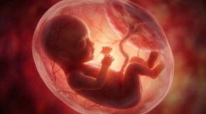
Cell transfer begins as early as during pregnancy. Some of the transferred cells are later broken down, while others appear to be able to remain in certain tissues. The direction of the transfer is also important: maternal cells can enter the child’s body. This maternal microchimerism is a distinct field of research and could, among other things, be significant for the development of the child’s immune system. Research therefore examines not only what the child leaves behind in the mother, but also how the mother’s cells might influence the child’s development.
Still Detectable Decades Later: Where Were the Cells Found?
Particularly surprising is the potentially long duration of the child’s cells’ presence. Scientists have detected cells of fetal origin in various maternal tissues, including bone marrow and other organs. A commonly used detection method relies on genetic differences between mother and child. In women who have given birth to a son, for example, researchers can search for cells that carry a Y chromosome. Since the mother herself does not normally possess a Y chromosome, this allows for the identification of a cell population of male origin in certain tests.
However, this approach has limitations: it primarily detects cells of male origin. Fetal cells from a pregnancy with a female fetus cannot be identified in the same way via a Y chromosome. An important early study by Bianchi and colleagues from 1996 detected male fetal cells in the blood of women whose pregnancies with sons had occurred many years earlier. Subsequent studies later investigated the detection of such cells in various tissues.
The results suggest that fetal microchimerism can persist long after a pregnancy. However, it is not yet fully understood how frequently certain cell types occur in individual organs or how long they survive there. Furthermore, the detection of a cell does not automatically prove that it performs a specific function in the organ in question.
Can Fetal Cells Help Repair Tissue?
One of the most intriguing questions concerns the possible function of the remaining cells. Some researchers suspect that certain fetal cells may be involved in repair processes. Evidence for this comes, among other sources, from studies in which fetal cells were detected in maternal tissues with injuries or pathological changes. The underlying hypothesis is that some of these cells may possess properties that enable them to respond to damaged tissue or participate in specific repair processes.
A review article by O’Donoghue (2008) described such observations and discussed the possibility that fetal cells might be involved in the response to tissue damage. However, the crucial question remains unanswered: Do these cells actually aid in healing, or do they merely accumulate at sites where damage already exists?
If fetal cells are found in injured tissue, this alone does not prove that they have improved the healing process. They could be performing an active function, reflecting an immune system response, or simply being present there without any significant functional role. Research must therefore investigate more closely which cell types are involved, what roles they play, and whether their presence actually leads to a measurable improvement in tissue function.
Between Protection and Risk: What Role Does the Immune System Play?
The mother’s body must perform a special immunological task during pregnancy. The child carries genetic traits from both parents and is therefore not completely genetically identical to the mother. Nevertheless, a pregnancy must normally be maintained without a harmful immune reaction. After birth, this biological peculiarity does not necessarily end completely. If fetal cells remain in the mother’s body, they can continue to interact with the immune system.
The consequences of this are an active area of research. There is evidence that microchimerism may be associated with both protective and adverse biological processes. For example, researchers are investigating whether certain cells are involved in tissue repair. At the same time, scientists are exploring possible links to autoimmune diseases, in which the immune system attacks the body’s own structures. A review article by Fugazzola, Cirello, and Beck-Peccoz, published in 2011 in Nature Reviews Endocrinology, summarized the findings on microchimerism and disease available at that time. The authors emphasized that the results are contradictory and that the role of fetal cells has not been clearly established.
The key limitation is this: A correlation between the presence of certain cells and a disease does not prove that these cells caused the disease. Nor is it proven that they generally protect against disease. It is also possible that the underlying disease itself influences how many cells enter a tissue or remain detectable there.
Is there a Link to Cancer or Other Diseases?
Possible relationships between fetal microchimerism and cancer are also being scientifically investigated. Fetal cells have been detected in tissue samples from tumors in various studies. This has raised the question of whether they might be involved in immune responses against tumor cells or whether, under certain conditions, they support processes that promote tumor growth. However, the findings to date do not present a consistent picture. The significance of these cells can vary depending on the tissue, cell type, disease, and biological context.
The review by Fugazzola and colleagues (2011) discusses both possible protective and potentially adverse associations. It shows that the mere presence of fetal cells in tumor tissue does not allow for a clear conclusion about their effect. It would therefore not be scientifically justified to portray fetal microchimerism as a protection against cancer or as a cause of cancer. For autoimmune diseases and cardiovascular diseases as well, the relationships have not yet been sufficiently clarified to derive concrete medical recommendations. The significance of microchimerism for the mother’s long-term health thus remains an open research question.
What Does this Mean for the Mother After Giving Birth?
For women who have already given birth, it is important to interpret these findings correctly. The detection of the child’s cells in the mother’s body is, first and foremost, a biological finding. It does not automatically mean that these cells confer a health benefit or pose a health risk. There are currently no established medical tests that would require routine screening for fetal microchimerism in healthy women. Likewise, there is no approved treatment specifically targeting these cells to improve health after pregnancy.
In the long term, research could help us better understand certain processes involved in tissue repair, immune regulation, or disease development. To achieve this, however, the cell types involved must be more precisely characterized, and their actual functions must be investigated. An important step would be to demonstrate that certain cells are not only present in a tissue but also perform a clearly defined function there, thereby causing a health-related effect. Until then, fetal microchimerism remains primarily a fascinating field of research and not a method for self-diagnosis.
Conclusion: A Biological Connection that Can Extend Beyond Pregnancy
Fetal microchimerism shows that the biological connection between mother and child is more complex than the exchange of nutrients, oxygen, and hormones via the placenta. Individual cells from the child can enter the mother’s body and, under certain conditions, remain detectable there for decades after pregnancy. Researchers are investigating whether these cells interact with the immune system, play a role in tissue repair, or are associated with certain diseases. However, it remains unclear what significance they actually have for the health of most women.
The key finding, therefore, is not that the baby biologically alters or protects the mother after birth. Rather, it appears that pregnancy can facilitate a long-lasting cellular exchange, the consequences of which scientists are only gradually beginning to understand. What sounds like a fascinating discovery today could provide new insights into immunobiology and tissue repair in the future. Whether this will lead to concrete medical applications remains to be seen in further studies.
Related Posts
-

When you are expecting, it seems that everyone around you has "valuable pregnancy and parenting…
-

DHA is one of the top recommended supplements for pregnant and nursing mothers. Docosahexaenoic acid…
-

Everyone's breast milk contains a unique collection of antibodies that are remarkably stable throughout breastfeeding…


