

The placenta could potentially provide doctors with earlier indications than before as to whether a pregnancy is at risk. Researchers have investigated whether additional information about the condition of the placenta can be obtained from routine ultrasound images. A computer-assisted analysis of the tissue structure could help identify pregnancies with an increased risk of adverse outcomes at an earlier stage.
In a recent study, scientists combined the analysis of ultrasound images with Doppler measurements and clinical data on the mother. The results suggest that additional examination of the placental structure could improve the assessment of pregnancy outcomes. The study was published in the journal “Placenta.”
More Information from Ultrasound
During prenatal care, the placenta is already routinely examined via ultrasound. Its blood flow can also be assessed using Doppler sonography, which reveals how blood flows through specific vessels. Changes in blood flow can, among other things, indicate that the unborn child’s blood supply may be compromised. However, Doppler primarily provides information about blood flow. The new method takes a different approach: the structure of the placental tissue. What initially appears on an ultrasound image as a pattern of light and dark areas—virtually impossible for laypeople to interpret—can be mathematically analyzed by a computer.
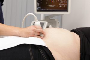
This involves what is known as image texture. Among other things, it describes how brightness values are distributed within the tissue and what patterns can be identified in the ultrasound image. Such characteristics could provide indirect clues about the structure of the placental tissue. The researchers therefore wanted to know whether this additional information could reveal anything about the subsequent course of the pregnancy.
What the Researchers Investigated
For the study, 129 pregnant women were examined. The ultrasound and Doppler measurements were taken at two different points during pregnancy. The scientists then evaluated various features of the ultrasound images and compared them with the course of the pregnancy and delivery. Of particular interest was the question of whether certain characteristics of the placental tissue were associated with the growth of the unborn child or with complications during pregnancy.
The analysis showed that certain features of placental texture were associated with the birth weight of the child. The Doppler measurements also provided information about the subsequent course of the pregnancy. It became particularly interesting when the researchers combined different data sets. A model that integrated clinical data, Doppler measurements, and computer-aided analysis of placental texture was better able to distinguish pregnancies with adverse fetal outcomes than models that considered only individual sources of information.
Which Complications Were the Focus
Among other things, the researchers considered severe preeclampsia, marked fetal growth restriction, and insufficient oxygen supply to be adverse pregnancy outcomes. In cases of fetal growth restriction, the baby grows more slowly than expected in the womb. One possible cause may be that the placenta is unable to adequately fulfill its role in supplying nutrients and oxygen. The baby may then not receive enough oxygen and nutrients.
The placenta can also play an important role in preeclampsia. The condition is characterized, among other things, by elevated blood pressure in the mother and can be dangerous for both mother and baby. Abnormal development or function of the placenta is associated with the biological processes that contribute to the condition.
Early detection of high-risk pregnancies could therefore be of great importance. The sooner abnormalities are detected, the more closely doctors can monitor further development and, if necessary, perform additional tests.
What the Ultrasound Pattern Might Reveal
The idea behind the new method is that changes in the placenta may be reflected not only in its blood flow but also in its tissue structure. The placenta consists of a complex network of tissue and numerous small blood vessels. This network changes continuously during pregnancy. At the same time, the placenta must adapt its performance to the increasing needs of the growing fetus.

Such changes are not necessarily visible to the naked eye on an ultrasound image. However, a computer can capture and compare significantly more image features simultaneously. This could reveal subtle differences that go unnoticed during a conventional visual assessment. The researchers therefore investigated whether certain features of the ultrasound images correlate with the actual structure of the placental tissue. In some cases, the imaging results were compared with microscopic examinations of the placental tissue after birth.
The results suggest that certain patterns in the ultrasound images may indeed contain information about the tissue’s fine structure. This is significant because changes within the placenta may occur even before clear clinical abnormalities become apparent.
Why the Combination of Different Tests Could Be Crucial
At the same time, the study suggests that no single measurement provides the decisive information. Rather, the combination of different data sets could be particularly valuable. Doppler imaging can provide clues about blood flow. Ultrasound analysis can offer additional information about tissue structure. Clinical data from the mother and measurements of the fetus complement this information.
Together, this creates a more comprehensive picture of the course of pregnancy. In the long term, such a combination could help assess risks more accurately than is possible with a single measurement.
This approach thus aligns with a general trend in medical research: existing examinations are evaluated using computer-aided methods so that additional information can be derived from the available data.
Could This Lead to a New Form of Prenatal Monitoring?
One advantage of the method would be that it would not necessarily require a completely new examination. Ultrasound is already a standard part of prenatal care. The additional computer-aided analysis could therefore, in principle, be based on existing ultrasound images.
For example, it is conceivable that software could automatically analyze certain characteristics of the placenta during or after an ultrasound examination. Combined with the results of the Doppler examination and other clinical data, this could lead to an additional risk assessment.
Such an application could be particularly valuable if it helps identify women at increased risk early on without the need for invasive tests. However, there is still a long way to go before such an application becomes a reality.
The Study Has Important Limitations
The study included only 129 pregnant women. This sample size is too small to draw reliable conclusions about how well the method would work in everyday clinical practice. The results must first be verified in larger studies. It would also be important to test the method on different patient groups and under various examination conditions.
It must also be clarified how reliably computer-aided image analysis works with ultrasound images from different devices. Ultrasound images can vary depending on the device, settings, examiner, and the baby’s position. A method intended for use in medical diagnostics in the future would need to be able to reliably account for these differences.
Furthermore, it must be investigated whether the method actually offers a practical advantage over already established examinations. A statistical correlation between certain image features and pregnancy outcomes does not automatically mean that the analysis can reliably predict complications.
Not Yet a New Prenatal Test
The results should therefore be viewed primarily as a first step. The study shows that computer-assisted analysis of the placental structure could provide additional information. However, it does not yet prove that this method can reliably predict which pregnancies will later develop complications.

For expectant parents, this means: The method is currently a research approach and not an available diagnostic test. Nor should the results be interpreted to mean that a specific abnormality in the placenta inevitably leads to a complication.
What is more interesting is the possibility that changes related to later pregnancy outcomes can be detected in the placenta at an early stage. The combination of image analysis, Doppler ultrasound, and clinical information could potentially be more accurate than evaluating individual measurements alone.
The Placenta as an Additional Window into Pregnancy Progression
If the results are confirmed in larger studies, analyzing placental structure could one day become another component of prenatal care. Doctors would then be able to assess not only the baby’s size or how blood flows through specific vessels, but also potentially detect more subtle changes within the placenta.
This could be particularly important in pregnancies with an increased risk of growth restriction, preeclampsia, or inadequate fetal growth. An earlier and more accurate assessment could, in turn, enable more targeted monitoring of the pregnancy.
It remains to be seen whether this idea will actually gain traction in medical practice. However, the current study provides an interesting indication that modern image analysis could reveal more from an ultrasound examination than is currently visible.
In the future, the placenta might thus be more than just an organ whose position, size, and blood flow are assessed during prenatal care. Its fine structure could potentially provide important information about how well a pregnancy is progressing—and whether closer monitoring might be necessary.
Related Posts
-

Researchers have found a mechanism in which mice lacking nutrients early in pregnancy have a…
-
The notion that mental illnesses originate exclusively in the brain is increasingly being challenged by…
-

Recent information shows that women who had their children after the age of 40 are…


